An introduction to electroencephalography (EEG)
Electroencephalography (EEG) is a technique used to measure electrical activity in the brain. This electrical activity is measured by electrodes on the brain and presented to the computer through an EEG amplifier. For about a century, EEG has been used in research to understand brain functioning or investigate diseases. More recently, these recordings have even been used to control computers, neuromarketing, and social interaction. In this blog, we will discuss the basics of EEG, how you can measure EEG and what you can do with EEG.

What is the origin of EEG signals?
The brain consists of millions of neurons. When recording EEG, not all brain activity from all structures can be measured or distinguished. EEG signals represent the summed electrical activity of populations of cortical neurons, meaning that activity from other structures in the brain may not be distinguishable in the EEG.1 The electrical signals, from action potentials, are the method of communication between neurons. When these action potentials travel along a neuron, a current dipole is generated (see image below). When many cortical neurons ‘fire’ action potentials synchronously (and are oriented in the same direction), electrodes on the scalp can measure this activity.
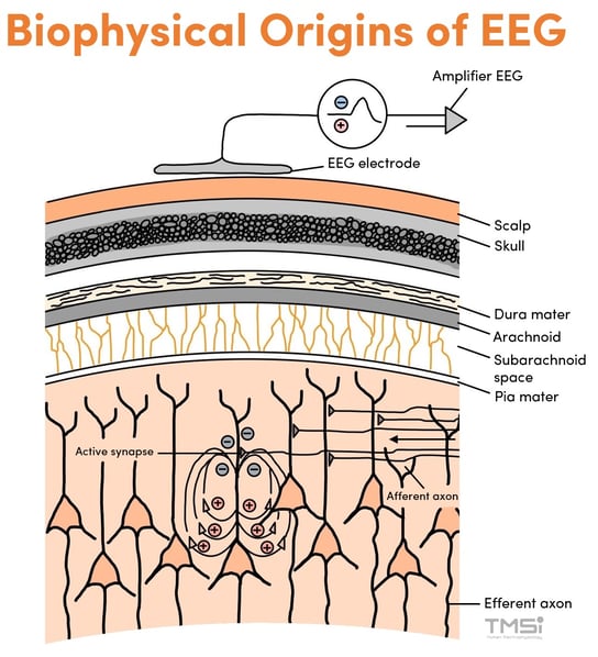 Origins of the EEG signal in the brain. Adapted from Bear et al1
The amplitudes of the brain waves measured from the scalp are usually in the range of 1 µV and 100 µV.2 Often, brain waves are irregular and no pattern can be recognized. However, sometimes distinct patterns can be recognized in the EEG, such as the four types of brain waves in the figure below.
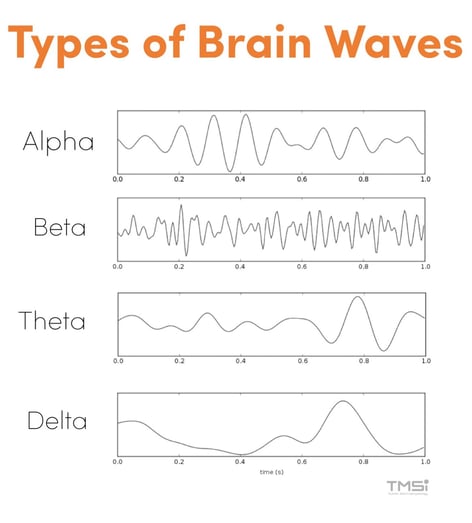
Type of brain waves (Picture source for alpha, beta, theta, and delta)
How can you measure EEG?
EEG Electrodes
EEG is measured non-invasively with electrodes on the scalp. There are different types of EEG electrodes on the market, such as gel, wet and dry electrodes. Each type has its advantages and disadvantages in terms of data quality and ease of use. The highest signal quality can be obtained using gel electrodes and are therefore the most commonly used. These electrodes are often integrated in a headcap that comes in different sizes. The placement of these electrodes is standardized and follows the 10-20 system for an even distribution of electrodes over the head.3
EEG Amplifier
The function of an EEG amplifier is to take the weak electrical signal at the scalp and to magnify its amplitude so that it can be further processed, recorded, or displayed on a computer. The EEG is always represented as a potential difference between recording sites, which is amplified. As a result, signals common to all electrodes are suppressed. For EEG there are different ways of referencing and amplifying a signal; an electrode can either be compared to one electrode (a common reference) or it can be compared to an averaged set of (all) electrodes.
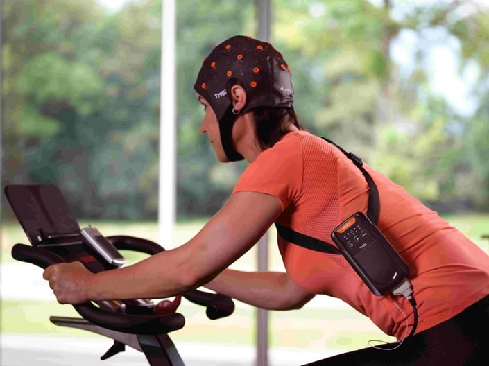
EEG amplifier and headcap
Considerations when recording EEG?
When done right, EEG can give us insights into brain functioning. However, there are multiple challenges to overcome to ensure you are measuring EEG accurately.
External EEG artifacts
As EEG is a biological potential with very low amplitude, it is highly sensitive to contamination from unwanted sources of interference.4 The most common external artifact (not originating from the body) that affects all electrodes is mains interference. This artifact is caused by environmental sources that surround the body and can be seen as 50 or 60 Hz noise.
Another common problem is cable movement artifacts. The cables between the electrode and the amplifier can be capacitively coupled to mains. A current can flow between the cables from one electrode to the other if this capacitive coupling is not the same for all cables. Using actively shielded cables, the capacitive coupling to mains is prevented and so 50/60 Hz noise is not introduced into the signal.
Physiological EEG artifacts
Artifacts originating from electrical activity produced by body parts other than the brain can sometimes be recorded at the scalp. For example, blinking causes a major artifact on the EEG as seen in the image below. Moreover, electrical activity caused by facial muscles like the jaw muscles are commonly present in the EEG. You can read more about how to recognize these physiological EEG artifacts here.
![]() Eye blink artifact in EEG
Eye blink artifact in EEG
What are applications of EEG?
EEG is used for many applications, such as social interaction, cognitive performance, and human machine interfacing.
To study social interaction, cognitive functions of multiple people are analyzed simultaneously while they perform a task. This is called hyperscanning.5 For hyperscanning, EEG should be recorded from the participants at the same time. The challenge is to ensure the data streams are synchronized perfectly.

Hyperscanning
Researchers or clinicians can assess the cognitive performance of subjects using event-related potentials. This is the neural response associated with a specific sensory, cognitive, or motor event and the timing of the response associated with this specific event. Research into cognitive performance can test the attention span, memory, and reaction time of participants and can give insight into neurological conditions.6
In Human Machine Interfacing with EEG, brain waves are used to control a computer or machine. This system should be able to acquire the EEG signal and extract features that are embedded in it. Based on this, the intention of the user can be understood so that electronic devices such as a PC, a robot, or a wheelchair can be controlled. This is also called a brain-computer interface (BCI).
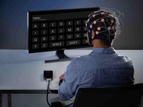
Brain-computer interface (BCI)
Conclusion
EEG is a very useful tool to gather insight into the brain activity. Using electrodes on the scalp and an EEG amplifier, brain waves can be recorded and interpreted by a researcher or clinician. By understanding measurement techniques and being aware of common artifacts, such as mains interferences or physiological artifacts, an accurate EEG signal can be measured. This can be used for many different applications in the fields of psychology, neuroscience, neurorehabilitation, and more.

References
1. Bear, M., Connors, B., & Paradiso, M. (2016). Neuroscience: Exploring the Brain (4th ed.). Wolters
Kluwer.
2. Webster J, Nimunkar A, Clark J. Medical instrumentation. 4th ed. 2010.
3. Klem GH, Lüders HO, Jasper HH, Elger C. The ten-twenty electrode system of the International Federation. The International Federation of Clinical Neurophysiology. Electroencephalogr Clin Neurophysiol Suppl. 1999;52:3-6.
4. Jiang X, Bian GB, Tian Z. Removal of Artifacts from EEG Signals: A Review. Sensors (Basel). 2019;19(5):987. Published 2019 Feb 26. doi:10.3390/s19050987
5. Czeszumski A, Eustergerling S, Lang A, et al. Hyperscanning: A Valid Method to Study Neural Inter-brain Underpinnings of Social Interaction. Front Hum Neurosci. 2020;14:39. Published 2020 Feb 28. doi:10.3389/fnhum.2020.00039
6. Woodman G. F. (2010). A brief introduction to the use of event-related potentials in studies of perception and attention. Attention, perception & psychophysics, 72(8), 2031–2046. https://doi.org/10.3758/APP.72.8.2031