A guide on how to recognize physiological artifacts
in the electroencephalogram (EEG) and how to manage them.
EEG artifacts are undesired signals that can introduce changes in the measurements and affect the signal of interest.1 As the EEG is a biological potential with very low amplitude (microvolts), it is highly sensitive to contamination from unwanted sources of interference. This contamination can be activity from electrophysiological sources other than the brain or from external sources from outside the body. In this blog, the most prevalent electrophysiological artifacts in the EEG are discussed. Moreover, this blog will describe how to recognize these artifacts on an EEG and discuss how to prevent or remove them.
What types of physiological artifacts can be seen on an EEG?
When measuring EEG with electrodes on the scalp, electrical potentials from extracerebral sources can contaminate the recording. These artifacts are hard to prevent and can distort the EEG in a way that, if not recognized, can mislead clinicians and cause a wrong interpretation of the EEG.2 For example, eye flutters may be wrongly identified as a representation of epilepsy (an interictal discharge), due to the similarities in representation.3
Types of EEG artifacts
The most common physiological signals found on an EEG are:
- Ocular activity or eye movements,
- Muscle activity,
- Cardiac activity.
What EEG artifacts are caused by eye movements?
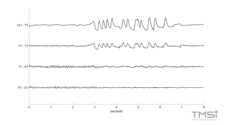
Eye blinks are a major source of contamination of EEG, with the greatest influence in (pre-)frontal electrodes, closest to the eyes. The amplitude of the blinking artifact is generally much larger (in the order of hundreds of microvolts) compared to that of the background EEG activity (a few to tens of microvolts).4 This artifact arises due to Bell’s Phenomenon: the eyeballs move in an upward direction as someone blinks. The eyeball has an electrical potential difference between the cornea (positively charged) and the retina (negatively charged). When the eyes roll upwards when blinking, this potential difference is changed and a positive waveform is produced in the electrodes closest to the eyes (frontal electrodes). When the eyes open, an opposite, negative waveform is seen in the EEG.

Figure 1: Blinking artifact on a few EEG traces. The blinking artifact with an amplitude of roughly 100-200 microvolts is mostly visible in the frontal electrodes.
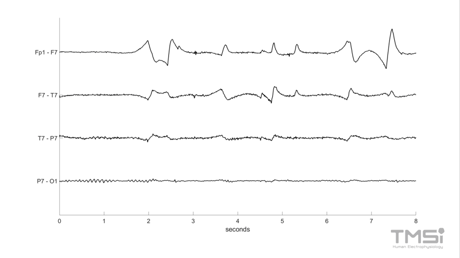
Lateral eye movements also produce waveforms in the electrodes closest to the temples (the F7 and F8 electrodes). For example, when looking to the right, the right cornea gets closer to the F8 electrode causing a positive wave and a negative wave at the F7 electrode.

Figure 2: Lateral eye movements artifact on a few EEG traces.
Eye movement artifacts are hard to prevent. The subject can be instructed to not blink and to look in one direction, but this can increase the cognitive load of a subject and is not always possible.5 By placing electrodes above, below, and lateral to the eye, the electrical activity caused by the movement of the eyeball (EOG) can be recorded in isolation. Post-processing algorithms like regression and blind-source separation can then be used to remove the artifacts.2 EEGlab is a useful interactive Matlab toolbox that can be used to analyze EEG data and perform e.g. blind-source separation techniques like the independent-component analysis.
What EEG artifacts are caused by muscle activity?
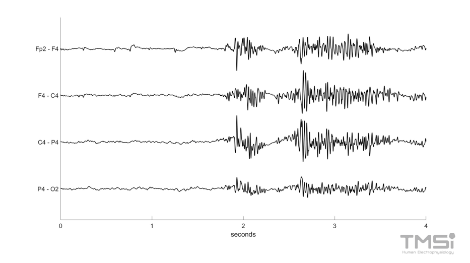
Muscles also produce electrical activity when they are contracted. When the head, face, or neck muscles are used, high-frequency (mostly > 30 Hz 6) signals are seen in most EEG electrodes. This generalized activity can be explained by volume conduction of muscular activity. The amplitude of this noise is dependent on the force of contraction and the type of muscle.7 Examples of movements that can produce artifacts are: chewing, tongue movements, or respiration.

Figure 3: Muscle activity artifacts on a few traces of EEG. This activity is produced by chewing.
Eliminating muscle artifacts from an EEG can be challenging. It is difficult to isolate the muscle activity from a single-channel measurement.1 Without this single-channel measurement, there are multiple approaches to limit the influence of muscle artifacts on EEG signals. They can be classified into filtering, linear regression, blind source separation, source decomposition, and neural networks.8
What EEG artifacts are caused by cardiac activity?
The electrical activity of the heart can be measured at the scalp with a low amplitude. Cardiac-related artifacts have a highly specific waveform (the QRS complex) and frequency. They are more likely to be present in the electrodes on the left side of the brain, as the heart is positioned in the left half of the chest. Whether this artifact is present, depends on the electrode positions and the body type of the subject. Removing this artifact can be done similarly to the eye movement artifact, as the cardiac activity can be measured separate from cerebral activity.9
Besides artifacts generated by direct activity from the heart, there are also pulse artifacts (‘cardioballistic artifact’) when an EEG electrode is placed over a pulsating vessel/artery. This can resemble EEG activity and is hard to correct.9
Conclusion
There are many artifacts that can show up on EEG recordings. To properly deal with artifacts, it is important to recognize the appearance of common artifacts and be able to trace them back to their origin in order to prevent, mediate, or filter them out. Besides physiological artifacts, EEG signals can also be affected by artifacts from external sources. If you want you can read more about common external artifacts here.
References
1. Jiang X, Bian GB, Tian Z. Removal of Artifacts from EEG Signals: A Review. Sensors (Basel). 2019;19(5):987. Published 2019 Feb 26. doi:10.3390/s19050987
2. Kaya İ. A Brief Summary of EEG Artifact Handling. Artif Intell. 2022. doi:10.5772/intechopen.99127
3. Mathias S, Bensalem-Owen M. Artifacts That Can Be Misinterpreted as Interictal Discharges. Journal of Clinical Neurophysiology. 2019;36(4):264-274. doi:10.1097/wnp.0000000000000605
4. Croft RJ, Barry RJ. Removal of ocular artifact from the EEG: a review. Neurophysiol Clin. 2000;30(1):5-19. doi:10.1016/S0987-7053(00)00055-1
5. Ochoa C, Polich J. P300 and blink instructions. Clinical Neurophysiology. 2000;111(1):93-98. doi:10.1016/s1388-2457(99)00209-6
6. Anderer P, Roberts S, Schlögl A et al. Artifact Processing in Computerized Analysis of Sleep EEG – A Review. Neuropsychobiology. 1999;40(3):150-157. doi:10.1159/000026613
7. McMenamin B, Shackman A, Maxwell J et al. Validation of ICA-based myogenic artifact correction for scalp and source-localized EEG. Neuroimage. 2010;49(3):2416-2432. doi:10.1016/j.neuroimage.2009.10.010
8. Minguillon J, Lopez-Gordo M, Pelayo F. Trends in EEG-BCI for daily-life: Requirements for artifact removal. Biomed Signal Process Control. 2017;31:407-418. doi:10.1016/j.bspc.2016.09.005
9. Urigüen J, Garcia-Zapirain B. EEG artifact removal—state-of-the-art and guidelines. J Neural Eng. 2015;12(3):031001. doi:10.1088/1741-2560/12/3/031001

