Discover insights into the benefits of high-density electromyography (HD-EMG), its distinctions from bipolar EMG, and the methodologies employed for measurement.
High-Density Electromyography (HD-EMG, also called high-density surface EMG or HDsEMG) measures both the spatial and temporal distribution of muscle activity, in terms of EMG amplitude, non-invasively on the skin’s surface. The EMG signals are acquired using at least four closely spaced electrodes from different locations over the muscle(s) of interest (1). In Figure 1, an example of an HD-EMG grid is included.
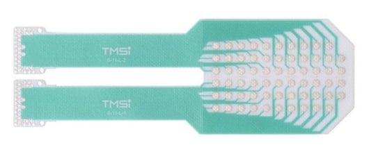 Figure 1: TMSi's Textile HD-EMG Grid (6x11 topology).
HD-EMG signals offer valuable insights into muscular activation, making it useful for neuromuscular research. The spatiotemporal signals provide information about regional activation of the muscles, including the magnitude and the size of the active region(s) of the muscle. The spatiotemporal recording characterizes how the action potential propagates along the muscle fiber, enabling estimation of muscle fiber properties such as average muscle fiber conduction velocity and innervation zone, the region where the alpha motor neuron attaches to the muscle fiber. Using decomposition methods, single motor unit activity can be extracted to represent the neural drive to the muscle. Examples of applications of HD-EMG are investigation of the effect of muscle fatigue and neuromuscular disorders (1).
What is the history of HD-EMG?
Increased computing capacity allowed for simultaneously recording of multiple EMG electrodes (2). In 1983, Masuda et al. (3) proposed the first electrode array of 16 stainless steel, strip electrodes to find the location of the neuromuscular junction at the biceps brachii (3, 4). Using these multi-electrode arrays, spatiotemporal information about muscle activation could be provided (2, 5). HD-EMG evolved from these multi-electrode arrays, offering 2D spatial data rather than just 1D spatial data. An example of an electrode array with stainless steel, strip electrodes is included in Figure 2.

Figure 2: Example of a grid surface electrode. From [6].
What is the origin of the HD-EMG signals?
HD-EMG signals are represented by bioelectricity, which refers to the potential differences measured on the skin's surface between two points within the muscle. The HD-EMG signals are composed of motor unit action potentials (MUAPs), which are the cumulative action potentials produced by all muscle fibers following the discharge of the innervating motor neuron. As the action potential travels through a muscle fiber, the 2D arrangement of the electrodes on the grid provides characteristics of muscle activity, such as single motor unit activity (7). More information about the origin of EMG signals is provided in this blog post about EMG.
What is the difference between bipolar EMG and HD-EMG?
Bipolar EMG is the conventional EMG method, measuring the difference in voltage between two electrodes on one muscle, representing muscle activity of that muscle. However, for some researchers, this provides insufficient information about muscle activity. The spatial resolution of HD-EMG is higher, with multiple channels evenly spaced in either one direction (array) or two directions (grid), as shown in Figure 3. The topographical distribution of electrodes allows for measurement of the activation of a large area of the muscle (8). Many variations on layouts of the grids exist. TMSi's grids can be reviewed in this article.
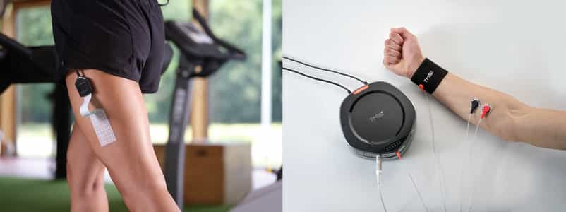
Figure 3: a) Textile HD-EMG grid (4x8L) b) Bipolar measurement of muscle activation, with 2 electrodes on the muscle and ground electrode on wristband.
Other than the number of electrodes, the electrode diameter of HD-EMG is smaller than conventional bipolar EMG. This reduction in electrode size aims to minimize the spatial low-pass filtering effect on the distribution of electric potentials on the skin (1). The low-pass filtering effects result from averaging the measured voltage beneath the surface of the electrode, which reduces the high-frequency content of the signal (9).
Lastly, the small inter-electrode distance (IED) of the HD-EMG grids increases the spatial resolution, facilitating signal interpolation for EMG images. The small IED guarantees the inclusion of all necessary muscle activation information needed for interpolation, preventing spatial aliasing (1, 7). Using an IED of max 10 mm gives an accurate image reconstruction (EMG image) (1). An example of EMG image reconstruction is included in Figure 4.
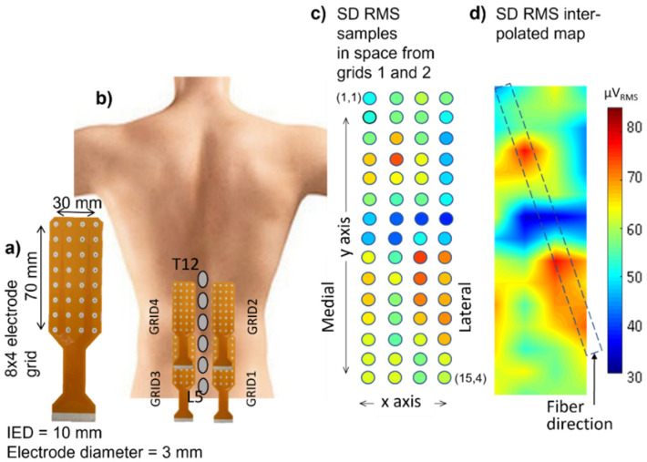
Figure 4 a) 8x4 electrode grid, b) Application of HD-EMG on each side of spine, c) Single Differential Root-Mean-Square Value, d) interpolated EMG image. From [9]
Conclusion
In summary, HD-EMG provides many valuable insights into the neuromuscular system, including the neural drive to the muscle, neuromuscular activation and muscle fiber characteristics.
TMSi’s Textile HD-EMG grids are available in different lay-outs, are flexible and form to the skin perfectly, avoiding movement artifacts. This makes it possible to study dynamic movements, such as walking or jumping, improving signal quality and reducing the need for artifact removal.
References
- Gallina, A., Disselhorst-Klug, C., Farina, D., Merletti, R., Besomi, M., Holobar, A., Enoka, R. M., Hug, F., Falla, D., Søgaard, K., McGill, K., Clancy, E. A., Carson, R. G., van Dieën, J. H., Gandevia, S., Lowery, M., Besier, T., Kiernan, M. C., Rothwell, J. C., … Hodges, P. W. (2022). Consensus for experimental design in electromyography (CEDE) project: High-density surface electromyography matrix. Journal of Electromyography and Kinesiology, 64, 102656. https://doi.org/10.1016/J.JELEKIN.2022.102656
- Avila, E. R., Williams, S. E., & Disselhorst-Klug, C. (2023). Advances in EMG measurement techniques, analysis procedures, and the impact of muscle mechanics on future requirements for the methodology. Journal of Biomechanics, 156, 111687. https://doi.org/10.1016/J.JBIOMECH.2023.111687
- Masuda, T., Miyano, H., & Sadoyama, T. (1983). The distribution of myoneural junctions in the biceps brachii investigated by surface electromyography. Electroencephalography and Clinical Neurophysiology, 56(6), 597–603. https://doi.org/10.1016/0013-4694(83)90027-5
- Merletti, R., Farina, D., & Gazzoni, M. (2003). The linear electrode array: a useful tool with many applications. Journal of Electromyography and Kinesiology, 13(1), 37–47. https://doi.org/10.1016/S1050-6411(02)00082-2
- Zwarts, M. J., & Stegeman, D. F. (2003). Multichannel surface EMG: Basic aspects and clinical utility. Muscle & Nerve, 28(1), 1–17. https://doi.org/10.1002/MUS.10358
- Masuda, T., & Sadoyama, T. (1991). Distribution of innervation zones in the human biceps brachii. Journal of Electromyography and Kinesiology, 1(2), 107–115. https://doi.org/10.1016/1050-6411(91)90004-O
- Merletti, R., Vieira, T. M., & Farina, D. (2016). Techniques for information extraction from the surface EMG signal: high-density surface EMG. In Surface Electromyography: Physiology, Engineering and Applications (pp. 126–157). Wiley-IEEE Press. https://doi.org/10.1002/9781119082934.CH05
- Besomi, M., Hodges, P. W., van Dieën, J., Carson, R. G., Clancy, E. A., Disselhorst-Klug, C., Holobar, A., Hug, F., Kiernan, M. C., Lowery, M., McGill, K., Merletti, R., Perreault, E., Søgaard, K., Tucker, K., Besier, T., Enoka, R., Falla, D., Farina, D., … Wrigley, T. (2019). Consensus for experimental design in electromyography (CEDE) project: Electrode selection matrix. Journal of Electromyography and Kinesiology, 48, 128–144. https://doi.org/10.1016/j.jelekin.2019.07.008
- Campanini, I., Merlo, A., Disselhorst-Klug, C., Mesin, L., Muceli, S., & Merletti, R. (2022). Fundamental Concepts of Bipolar and High-Density Surface EMG Understanding and Teaching for Clinical, Occupational, and Sport Applications: Origin, Detection, and Main Errors. Sensors, 22(11), 4150. https://doi.org/10.3390/S22114150/S1
- del Vecchio, A., Holobar, A., Falla, D., Felici, F., Enoka, R. M., & Farina, D. (2020). Tutorial: Analysis of motor unit discharge characteristics from high-density surface EMG signals. Journal of Electromyography and Kinesiology, 53, 102426. https://doi.org/10.1016/J.JELEKIN.2020.102426
- Rojas-Martínez, M., Serna, L. Y., Jordanic, M., Marateb, H. R., Merletti, R., & Mañanas, M. Á. (2020). High-density surface electromyography signals during isometric contractions of elbow muscles of healthy humans. Scientific Data 2020 7:1, 7(1), 1–12. https://doi.org/10.1038/s41597-020-00717-6
- Merletti, R., & Muceli, S. (2019). Tutorial. Surface EMG detection in space and time: Best practices. Journal of Electromyography and Kinesiology, 49, 102363. https://doi.org/10.1016/J.JELEKIN.2019.102363